Is apparent tarsal vascular rarefaction a companion marker of meibomian gland dropout?
Is apparent tarsal vascular rarefaction a companion marker of meibomian gland dropout?
May 12, 2026
Ductal obstruction, altered meibum, and gland atrophy remain central to current models of meibomian gland dysfunction. Lid-margin vascularity and vascular engorgement are also recognised features in MGD assessment.
However, less attention appears to have been given to the vascular network within the tarsal region itself.
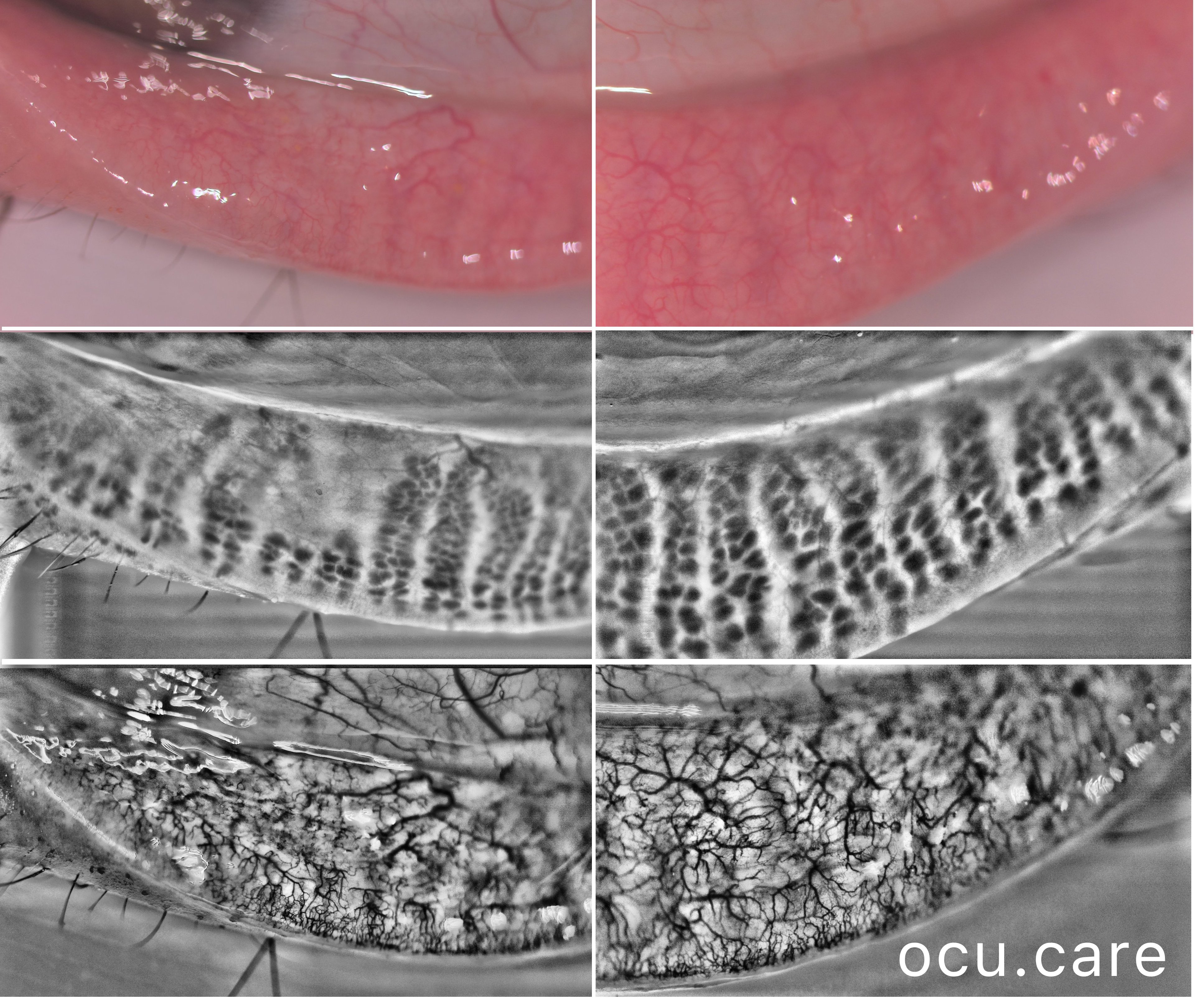
In the presented images, we show two contrasting examples: one region where meibomian gland dropout appears to coincide with reduced visible vascular network density, and another region with preserved gland architecture and a denser visible vascular network.
This observation is particularly interesting in light of Obata’s histopathological work, which described vascular structures around meibomian gland acini and highlighted the potential relevance of local blood supply to lipogenesis and gland function.
While obstruction and altered meibum remain central to MGD pathogenesis, the relationship between gland dropout and the surrounding tarsal vascular network may deserve closer investigation.
Reference: Obata H. Anatomy and Histopathology of Human Meibomian Gland. Cornea. 2002;21 Suppl 2:S70–S74.