When the gland is still visible — but the architecture is already changing
Jun 2, 2026
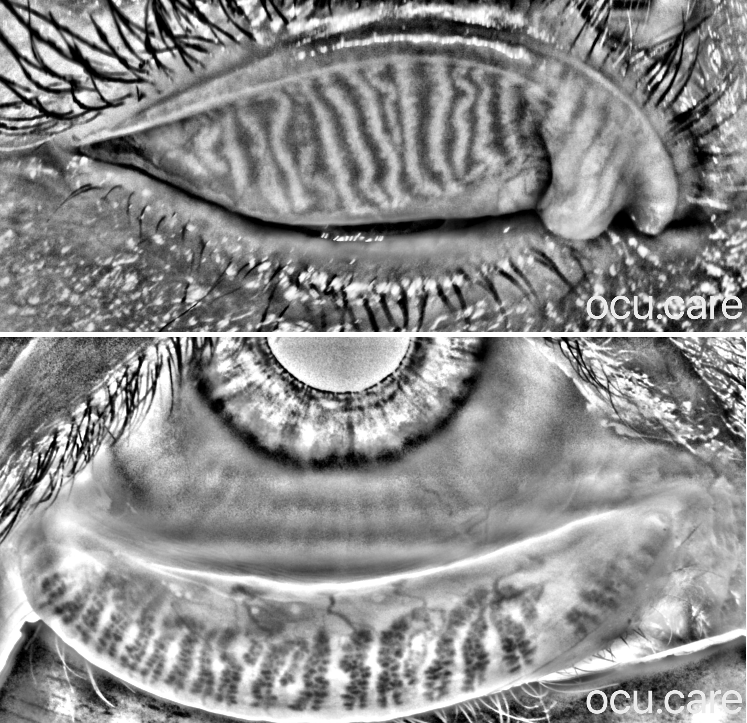
Upper and lower lid meibography in a 63-year-old woman shows why meibography should not be interpreted only as a “dropout score.”
The lower eyelid shows telangiectasia, with fragmented and shortened meibomian glands, more pronounced nasally.
The upper eyelid shows a different pattern: mild tortuosity, fragmented gland architecture, and fluffy-appearing areas, more evident nasally than temporally.
These fluffy areas are clinically interesting because they may represent stagnant meibum, acinar dilation, altered gland architecture, or a transitional stage before gland loss.
For patients, meibography is a powerful educational tool. By visualising the underlying gland changes, it helps them understand why early and consistent management is important.
Repeat imaging allows us to compare findings over time and assess whether the glands remain stable, continue to deteriorate, or show signs of improvement with treatment.